The work in our laboratory is possible thanks to funding from the National Institute on Deafness and Other Communication Disorders (NIH R01 DC017708) and models from the Aged Rodent Colonies at the National Institute on Aging.
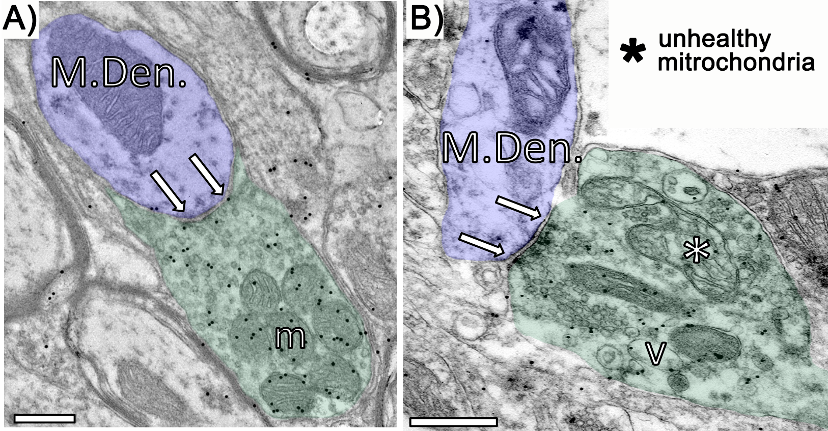
What ultrastructural changes are occurring to GABAergic synapses in the auditory midbrain before age-related hearing loss occurs? It has been well documented that GABA is downregulated in the inferior colliculus with age. While the effects of this lost GABA are becoming understood at older ages, we have very little information regarding what GABAergic changes occur during middle ages when hearing is typically unaffected by aging. We are seeking to determine whether age-related changes to GABAergic synapses are occurring before deficits to temporal processing and hearing thresholds. To answer these questions we use a number of techniques (immunogold transmission electron microscopy, prepulse inhibition of the acoustic startle reflex, envelope frequency following responses, auditory brainstem response) across several age groups: To the right is an electron micrograph showing a young GABAergic synapse (bracketed by the arrows; green = presynaptic bouton; blue = postsynaptic target) onto medium sized dendrites at young (A) and old (B). In aged tissue, we also find that mitochondrial cristae are often swollen and that the mitochondria are distorted and appear unhealthy. |
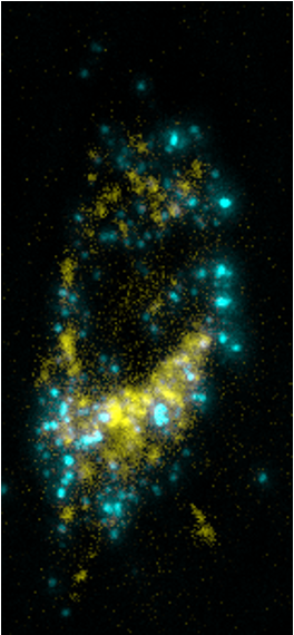
GABAergic synapses in the inferior colliculus are lost with age, but what circuit(s) is actually losing this input? The inferior colliculus is often referred to the auditory nexus or hub as it 1) receives ascending input from over a dozen nuclei, 2) is the origin of glutamatergic and GABAergic projections that target a number of ascending targets and descending targets and 3) receives massive descending inputs from the cortex and non-lemniscal auditory thalamus. We are determining whether descending and/or ascending IC circuits are losing GABAergic input during aging. To do this we are combining a intersectional viral strategy with immunogold transmission electron microscopy. To the left is a cell in the lemniscal region of the inferior colliculus expressing GFP after a deposit of CAV2 in the auditory thalamus and AAV in the inferior colliculus; the inset demonstrates the value of such viral techniques as spines are also well labeled. |
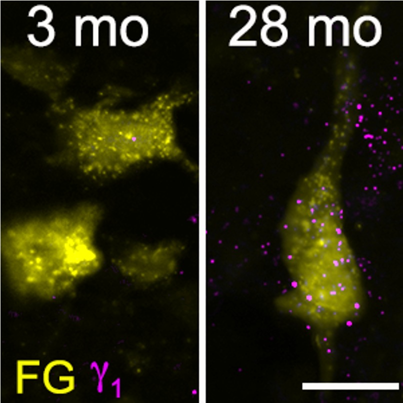
Which IC circuits express the GABAAγ1 subunit before the onset of auditory processing deficits. Our lab’s first publication demonstrated that the GABAAγ1 subunit is maximally expressed during middle age on both GABAergic and presumptive glutamatergic cells in the inferior colliculus. The GABAAγ1 subunit, which is virtually absent during young ages, is increasingly expressed with age and may have dramatic effects on the physiology of IC cells as this subunit has been shown to increase GABA efficacy by increasing the receptor’s permeability to chloride (Caspary et al., 1999; Milbrandt et. al., 1997; Wafford et al., 1993).We are seeking to determine whether ascending and/descending IC circuits express the GABAAγ1 subunit before deficits to temporal processing and hearing thresholds. We will use fluorescent tract-tracing, immunohistochemistry for the GABAAγ1 subunit and GAD, prepulse inhibition of the acoustic startle reflex, envelope frequency following responses, auditory brainstem response across our age groups. To the right are young and old FluoroGold labeled IC cells that project to the auditory thalamus. Note the higher expression of the GABAAγ1 subunit at 28 months than at 3 months |
We discovered that perineuronal nets (PN) are upregulated with age in the inferior colliculus. PNs are known for their multifaceted roles in development and aging (e.g. reducing structural plasticity and facilitating synaptic plasticity). Most recently we examined this increase of PNs with tract-tracing the inferior colliculus-auditory thalamus pathway. We discovered that PNs increase on non-lemniscal IC pathways during middle age while not increasing in the lemniscal IC until old age. We are currently pursuing identifying PN expression at an ultrastructural level to determine their relationship with aging synaptic arrangements. Below is a fluorescent micrograph showing two netted aged IC cells (one GAD-positive, arrow; one GAD-negative, arrowhead) that project to the auditory thalamus.
|